Molecular imaging technique could improve breast cancer screening
28 Mar 2024
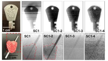
Cancer detection Mammographic images (A, B) of lesions in the left breast, confirmed as invasive ductal carcinoma (arrow) and fibroadenoma (double arrow). Left PEM images (C, D) show intense uptake in the known cancer and no uptake in the fibroadenoma. Right PEM images (E, F) do not show any abnormal uptake. Left PEM images obtained 1 h (G) and 4 h (H) after 18F-FDG injection show no substantial visual difference in uptake. (Courtesy: Radiological Society of North America)
Mammography is a widely employed and effective tool for early detection of breast cancer, but dense
You’ve reached the limit of what you can view on Physics World without registering
If you already have an account on Physics World, then please sign in to continue reading
If you do not yet have an account, please register so you can
Access more than 20 years of online content
Choose which e-mail newsletters you would like to receive
Cynthia E Keen is a freelance journalist specializing in medicine and healthcare-related innovations